Curious Mummies exhibition: multimedia content creation
The exhibition ‘Curious Mummies: From the Champollions to the…

Home » Articles and News » Promotion of the collections of the French National Museum of Natural History
The e-col+ project, led by the National Museum of Natural History, aims to showcase France’s natural history collections through digitisation. By establishing 3D digitisation workflows and developing artificial intelligence tools, the project enables researchers to utilize and enrich these resources.
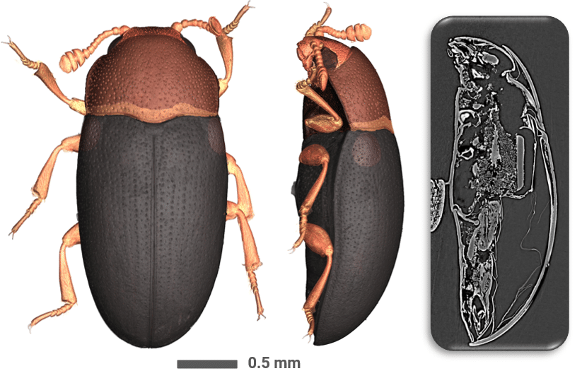
NOVITOM was selected to provide outsourced microtomography services. High-resolution scans of an entomological collection were performed at the Grenoble ESRF synchrotron, using the BM05 and BM18 instruments.
What makes this project unique is the large number of specimens to be scanned, as well as their fragility.
NOVITOM has developed a specific protocol for analysing microtomographic data. Once the samples have been received, the first step involves planning and carrying out the acquisition of synchrotron microtomography data, as well as the reconstruction of that data.
A 3D U-Net architecture is trained to segment the annotated data generated using NOVITOM’s manual labelling software. A mesh is then created. To do this, the segmented data is dilated to fill in small gaps, then the dilated volume is converted into a 3D mesh, and finally the complexity of the mesh is reduced whilst retaining its overall shape. This lightweight final rendering makes it easy to share the results obtained.
The data analysis protocol was presented at the Technart2025 international conference, which took place in Perugia (Italy).
Le projet « e-COL+ Valorisation des données naturalistes » is supported by a grant from the French government managed by the ‘ANR, the French National Research Agency (ANR-21-ESRE-0053 PIA3)
As an independent laboratory, Novitom enables organisations of all kinds to access synchrotron micro- and nano-tomography.
>>Our services and expertise in natural and cultural heritage
The exhibition ‘Curious Mummies: From the Champollions to the…
The e-col+ project, led by the National Museum of…
X-ray tomography is now a reference method for non-destructive…
3D digitisation enables the creation of a detailed digital representation of specimens in natural history collections, in particular through X-ray microtomography. It facilitates their study, analysis and promotion without the need to handle the specimens directly.
Synchrotron microtomography provides information on the internal structure of delicate specimens in a non-invasive manner. This method therefore allows microstructures to be studied without damaging the samples.
Microtomography can be adapted to projects involving a large number of specimens, typically several hundreds.
Microtomographic data enable the internal structures of the specimen to be explored (such as organ-related segmentation) and various quantitative details to be extracted: volumes, surface areas, thicknesses and other specific morphological features.
Yes. In this project, Novitom has developed a protocol for analysing 3D images using artificial intelligence. In particular, a 3D U-Net architecture is trained to segment the annotated data and automatically isolate certain structures of interest.
3D segmentation involves identifying and isolating, within the volumes produced by microtomography, the structures or elements of interest for analysis. It then enables measurements to be taken and 3D representations to be generated that can be used by researchers or the general public.
Yes. After segmentation, the data can be converted into a 3D mesh. This can then be simplified to preserve the overall shape of the specimen whilst producing a smaller file that is easier to share. These models are also used to produce replicas via additive manufacturing.