Tracking the evolution of materials microstructure : in situ mechanical testing
Why perform microCT during tensile/compression tests? The use of…
Home » Our services » Multi-scale tomography » AI-powered 3D image analysis
X-ray tomography (CT, micro-CT) generates 3D image volumes that are extremely rich in information. However, extracting quantitative data can be complex when contrast is low or structures are interlaced.
Artificial intelligence methods based on deep learning, and in particular convolutional neural networks (CNNs), now make it possible to push the boundaries of conventional approaches and automate the analysis of tomography volumes with high accuracy and reproducibility.
Thanks to its pre-trained U-Net 3D network, NOVITOM provides advanced and robust image analysis services to efficiently process your volumes from micro-tomography, nano-tomography or industrial tomography.
Segmentation of complex or ambiguous structures: proportion of total volume occupied by pores
Size and shape distribution: equivalent diameter, average size, sphericity, shape factor, orientation, Feret diameter.
Spatial location: position of pores within the component or structure.
Connectivity: distinction between open and closed pores, dimensions of the interconnected pore network, local thickness, tortuosity.
Dynamic evolution of porosity under mechanical, thermal or chemical stresses
Foam cells: cell volume, cell density, size distribution
Â
Â
Yes, the AI-specific algorithms we have developed deliver consistent and standardised results, regardless of the operator. This ensures the reproducibility of the analysis.
Yes. Our specially trained algorithms make it possible to detect all internal defects present in the images. By applying criteria to the defects identified, we achieve a reduction in false negatives and false positives.
Consistent, standardised results, independent of the operator.
Detection of all internal defects in 3D, reduction in false negatives and false positives
Automated analysis of batches or series of 3D volumes with drastically reducing processing time
Details within complex, multi-material objects and assembly, multi-scale analysis, irregular objects and interfaces
Efficient correction of beam-hardening artefacts, noise, super-resolution, etc.
Why perform microCT during tensile/compression tests? The use of…
A recent study conducted by Xploraytion, Novitom, Merck and…
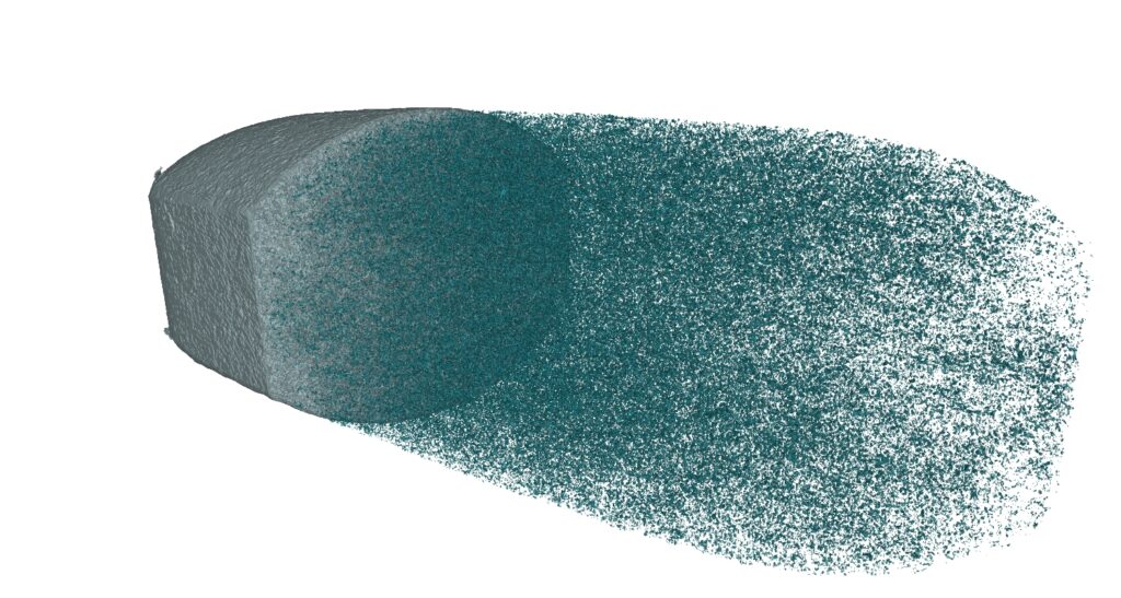
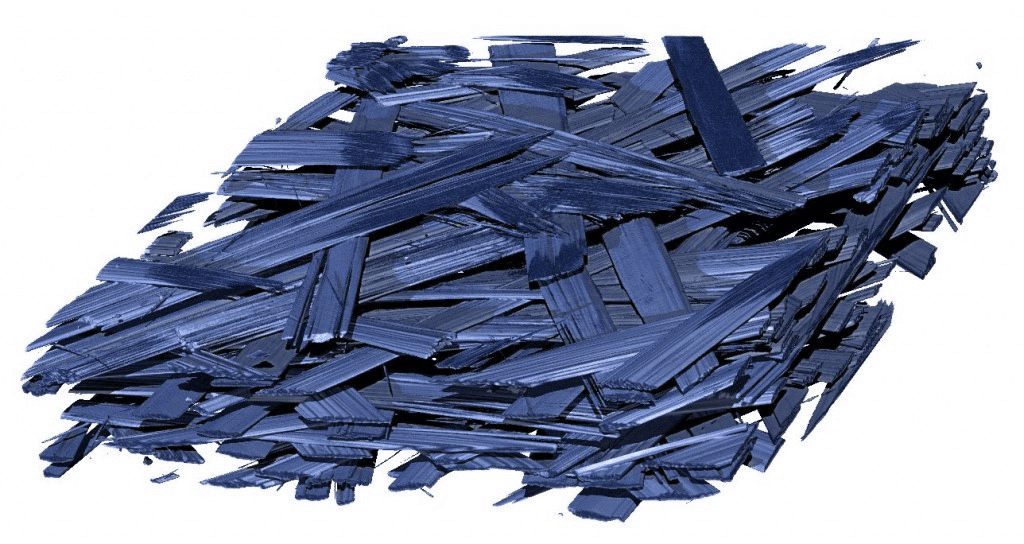
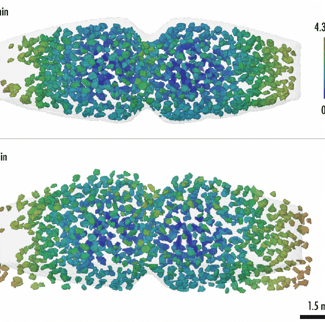
The images from X-ray microtomography (μCT) allow for advanced…
For measuring both R&D samples and finished products:
For visualisations and quantitative results tailored to your needs:
thanks to the use of a single pre-trained CNN network, specifically designed for analysing CT images
tailored to your requirements, with a technical contact person who supports you from start to finish
thanks to detailed reports and 3D visualisations that facilitate decision-making