Cosmetics

An AFM image of skin collagen showing 16 sub-images for subsequent scoring using a pre-trained AI algorithm.

Quantifying dermal collagen organisation with AI-assisted AFM

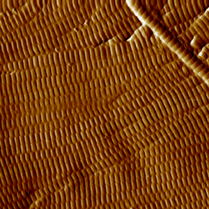
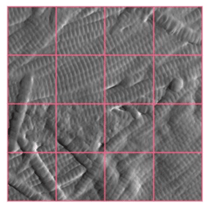
A new approach for assessing skin ageing and treatment efficacy Understanding how collagen organisation changes with age remains a major challenge in skin research. While macroscopic alterations can be identified at the fibre bundle scale using conventional histology, many of the most significant age-related modifications occur at a much finer

More »

Assessment of strengthening the mechanical properties of hair fibers

Atomic force microscopy (AFM) is used to map the topology (i.e. profilometry) of the cuticle while measuring the mechanical properties of adhesion and Young’s modulus in the same area, with nanometric measurement steps. These measurements are used to assess the quality of the cuticle and its adhesion properties. Images obtained

More »
volume tomographique révélant d'intérieur d'une montre

Why choose X-ray tomography or microCT?

X-ray tomography is now a reference method for non-destructive three-dimensional imaging. Its principle is based on capturing a large number of X-ray images from different angles, which are then combined digitally to reconstruct the internal structure of an object. Tomography, which has become essential in various fields such as material

More »
dessin illustrant la structure du stratum corneum et le dépôt d'un produit cosmétique en surface

Unveiling the impact of topical formulations on the organisation of skin lipid barrier

X-ray diffraction reveals the structural organisation of the intercorneocyte lipids in the stratum corneum X-ray microdiffraction (µXRD) unveils the structural organization of the intercorneocyte lipids in the stratum corneum : Lamellar stacking (SAXS) => layer thickness and organization quality Crystalline and fluid structures inside the lipid layers (WAXS) => hexagonal/orthorhombic/amorphous

More »
visualisation 3D de l'orientation des fibres dans un polymère composite par tomographie RX

3D characterisation of fibres orientation in a composite

The images from X-ray microtomography (μCT) allow for advanced analyses at the fiber level: composites, fabrics, plants, paper. We have developed algorithms dedicated to high-resolution image analyses in order to calculate the orientation fields of fibers in samples or complete parts. The material is probed ‘as-is’, without the need to

More »
Volume 3D colorisé illustrant la segmentation des phases et grains dans du ciment.

Why choose microCT to study particle size?

X-ray microtomography (micro-CT) images enable advanced particle size analysis, allowing the characterisation of the size, shape, and spatial distribution of particles, pores, or inclusions that make up one or more phases of a material. The material is probed “as is” , i.e. without the need for conditioning. The three-dimensional nature

More »
image obtenue par AFM d'une coupe de peau : les fibres de collagène bien organisées sont visibles

Anti-aging skincare seen through collagen fibres organisation

Novitom has developed an original test based on the analysis of the quality of the organisation of collagen fibres. Collagen, which is the main constituent of the dermis, largely determines its flexibility during aging. Two techniques are combined: Atomic force microscopy (AFM) provides spectacular images of the fibres in varying

More »
montage en couleur révélant des signaux de diffraction RX

Structural analysis of human Stratum corneum lamellar structure

Congratulations to our colleague Danilo Rosa Nunes, PhD in Pharmaceutical Science! He won thes Young Researcher Award following the competition organised as part of the Lipids and Cosmetics 2022 conference. The awarded work is described in a poster: ”Quantitative structural analysis of Human Stratum Corneum lamellar structure by Computational Simulation of

More »