Biomedical

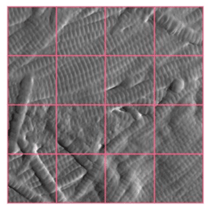
An AFM image of skin collagen showing 16 sub-images for subsequent scoring using a pre-trained AI algorithm.

Quantifying dermal collagen organisation with AI-assisted AFM

A new approach for assessing skin ageing and treatment efficacy Understanding how collagen organisation changes with age remains a major challenge in skin research. While macroscopic alterations can be identified at the fibre bundle scale using conventional histology, many of the most significant age-related modifications occur at a much finer

More »
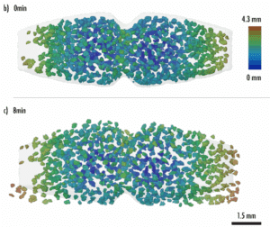
principe actif rendu visible en 3D par microCT au cours de la dissolution d'un comprimé

The microstructure of tablets revealed by synchrotron X-rays

A recent study conducted by Xploraytion, Novitom, Merck and their partners shows how the internal structures of tablets are visualised in 3D, non-destructively and in real time. The resolution, speed, and sensitivity of synchrotron X-rays make it possible to capture coating uniformity, porosity, and disintegration dynamics in just a few

More »
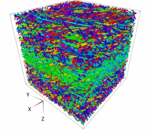
visualisation 3D de l'orientation des fibres dans un polymère composite par tomographie RX

3D characterisation of fibres orientation in a composite

The images from X-ray microtomography (μCT) allow for advanced analyses at the fiber level: composites, fabrics, plants, paper. We have developed algorithms dedicated to high-resolution image analyses in order to calculate the orientation fields of fibers in samples or complete parts. The material is probed ‘as-is’, without the need to

More »
Volume 3D colorisé illustrant la segmentation des phases et grains dans du ciment.

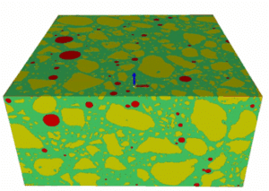
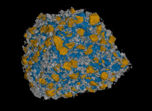
Why choose microCT to study particle size?

Though X-ray microtomography (micro-CT), Novitom provides advanced particle size analysis, allowing the characterisation of the size, shape, and spatial distribution of particles, pores, or inclusions that make up one or more phases of a material. The material is probed “as is” , i.e. without the need for conditioning. The three-dimensional

More »
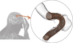
Image illustrant un anévrisme cérébral avec un dispositif médical de type stent à maille sérée inséré pour traitement par embolisation.

From patient specific aneurysm data to model for computational fluid dynamics

In a study on patient-specific modeling of blood flow in cerebral aneurysms, Novitom played a key role by providing high-resolution synchrotron X-ray microtomography of in vitro aneurysm models treated with endovascular coils. Using monochromatic radiation at the European Synchrotron Radiation Facility (ESRF), Novitom acquired and segmented 3D datasets at micrometer

More »
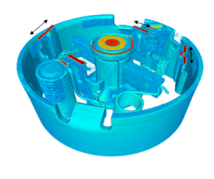
repésentation 3D des écarts géométriques entre la pièce dessinée et la pièce contrôlée

Comparison of the scanned full volume of a part with its CAD model

High-resolution, high-energy X-ray tomography provides non-destructive access to the entire part, including internal areas that are inaccessible to other techniques. NOVITOM has developed an algorithm for comparing the CAD part with the volume reconstructed by tomography obtained on the manufactured part. The image next to this text is a 3D

More »

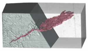
3D localisation and visualisation of cracks

Microtomography enables the non-destructive detection and 3D visualization of cracks. It accurately quantifies the size, shape, and volume of cracks, including their propagation depth. This volumetric measurement capability allows for the assessment of structural integrity, the monitoring of degradation progression, and the optimization of materials. Essential in R&D, it helps

More »
volume tomographique révélant d'intérieur d'une montre

Why choose X-ray tomography or microCT?

X-ray tomography is now a reference method for non-destructive three-dimensional investigation of materials and objects. Its principle is based on capturing a large number of X-ray images from different angles, which are then combined digitally to reconstruct the internal structure of an object. Tomography, which has become essential in various

More »
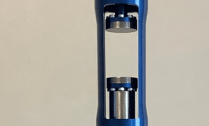
configuration expérimentale en diffraction RX haut débit ESRF

Crystalline structural analysis with high-throughput X-ray powder diffraction

We are enthusiastic to announce the first industrial use by Novitom of the 𝗮𝘂𝘁𝗼𝗺𝗮𝘁𝗲𝗱, 𝗵𝗶𝗴𝗵-𝘁𝗵𝗿𝗼𝘂𝗴𝗵𝗽𝘂𝘁 𝗫-𝗿𝗮𝘆 𝗽𝗼𝘄𝗱𝗲𝗿 𝗱𝗶𝗳𝗳𝗿𝗮𝗰𝘁𝗶𝗼𝗻 (𝗫𝗥𝗣𝗗) 𝘀𝘆𝘀𝘁𝗲𝗺 at 𝗘𝗦𝗥𝗙. Further, we have used it for non-standard powders as samples were metals’ sheets ! X-ray diffraction provides valuable insights into the crystalline structure, phase composition, and purity of

More »