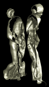
Promotion of the collections of the French National Museum of Natural History
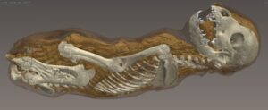
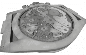
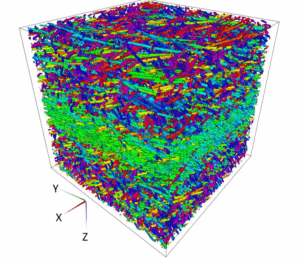
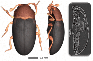
The e-col+ project, led by the National Museum of Natural History, aims to showcase France’s natural history collections through digitisation. By establishing 3D digitisation workflows and developing artificial intelligence tools, the project enables researchers to utilize and enrich these resources. NOVITOM was selected to provide outsourced microtomography services. High-resolution scans