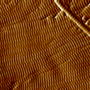
Assessment of strengthening the mechanical properties of hair fibers

Atomic force microscopy (AFM) is used to map the topology (i.e. profilometry) of the cuticle while measuring the mechanical properties of adhesion and Young’s modulus in the same area, with nanometric measurement steps. These measurements are used to assess the quality of the cuticle and its adhesion properties. Images obtained

More »
repésentation 3D des écarts géométriques entre la pièce dessinée et la pièce contrôlée

Comparison of the scanned full volume of a part with its CAD model

High-resolution, high-energy X-ray tomography provides non-destructive access to the entire part, including internal areas that are inaccessible to other techniques. NOVITOM has developed an algorithm for comparing the CAD part with the volume reconstructed by tomography obtained on the manufactured part. The image next to this text is a 3D

More »

3D localisation and visualisation of cracks

Microtomography enables the non-destructive detection and 3D visualization of cracks. It accurately quantifies the size, shape, and volume of cracks, including their propagation depth. This volumetric measurement capability allows for the assessment of structural integrity, the monitoring of degradation progression, and the optimization of materials. Essential in R&D, it helps

More »
volume tomographique révélant d'intérieur d'une montre

Why choose X-ray tomography or microCT?

X-ray tomography is now a reference method for non-destructive three-dimensional investigation of materials and objects. Its principle is based on capturing a large number of X-ray images from different angles, which are then combined digitally to reconstruct the internal structure of an object. Tomography, which has become essential in various

More »
virtual cutting of a statuette after X-ray tomography of the entire piece

Searching for clues to authenticate a statuette

Searching for clues leading to the authentication of a work of art is a complex process that requires consideration of all aspects of the work, including artistic and historical aspects, of course, but also the materials used. For statues, the difficulty lies in the fact that the interior is not

More »
configuration expérimentale en diffraction RX haut débit ESRF

Crystalline structural analysis with high-throughput X-ray powder diffraction

We are enthusiastic to announce the first industrial use by Novitom of the 𝗮𝘂𝘁𝗼𝗺𝗮𝘁𝗲𝗱, 𝗵𝗶𝗴𝗵-𝘁𝗵𝗿𝗼𝘂𝗴𝗵𝗽𝘂𝘁 𝗫-𝗿𝗮𝘆 𝗽𝗼𝘄𝗱𝗲𝗿 𝗱𝗶𝗳𝗳𝗿𝗮𝗰𝘁𝗶𝗼𝗻 (𝗫𝗥𝗣𝗗) 𝘀𝘆𝘀𝘁𝗲𝗺 at 𝗘𝗦𝗥𝗙. Further, we have used it for non-standard powders as samples were metals’ sheets ! X-ray diffraction provides valuable insights into the crystalline structure, phase composition, and purity of

More »
extrait de webinaire décrivant l'insertion de défauts virtuels via le logiciel de simulation CT Novi-Sim

Optimizing X-ray CT with virtual tomography

On September 13, 2023, Rigaku hosted an episode of its Ask the Expert webinar series, featuring Dr. Awen Autret, R&D Engineer at Novitom. The session, titled “Virtual Tomography – Optimizing Data Acquisition Parameters Without a CT Scanner”, explored how advanced simulation can accelerate and enhance X-ray CT imaging workflows. Dr.

More »
image obtenue par AFM d'une coupe de peau : les fibres de collagène bien organisées sont visibles

Anti-aging skincare seen through collagen fibres organisation

Novitom has developed an original test based on the analysis of the quality of the organisation of collagen fibres. Collagen, which is the main constituent of the dermis, largely determines its flexibility during aging. Two techniques are combined: Atomic force microscopy (AFM) provides spectacular images of the fibres in varying

More »

International Conference on Industrial Computed Tomography (iCT2023)

Novitom and Xploraytion attended the 12th International Conference on Industrial Computed Tomography (iCT2023) that took place in March 2023 Fürth Germany. We were delighted to present recent work with an oral presentation and a poster. And we enjoyed lively discussions with the participants! Novi-Sim is a X ray radiography and

More »