Home » Technical means » X-ray tomography » Synchrotron nanotomography
Synchrotron nanotomography (SR-nano-CT) is the high-resolution version of synchrotron microtomography, a non-destructive 3D imaging technique.
It is particularly well-suited for studying materials organised at the submicrometer scale, and for characterising porosities, fibres, inclusions, interfaces, and biological tissues at scales inaccessible to conventional non-destructive techniques.
Visualisation of submicron structures in 3D
High contrast, including between weakly absorbing materials
Possible coupling with stimulation devices for 4D studies: growth, corrosion, cracking, etc.
Non-destructive analysis of fragile or rare samples
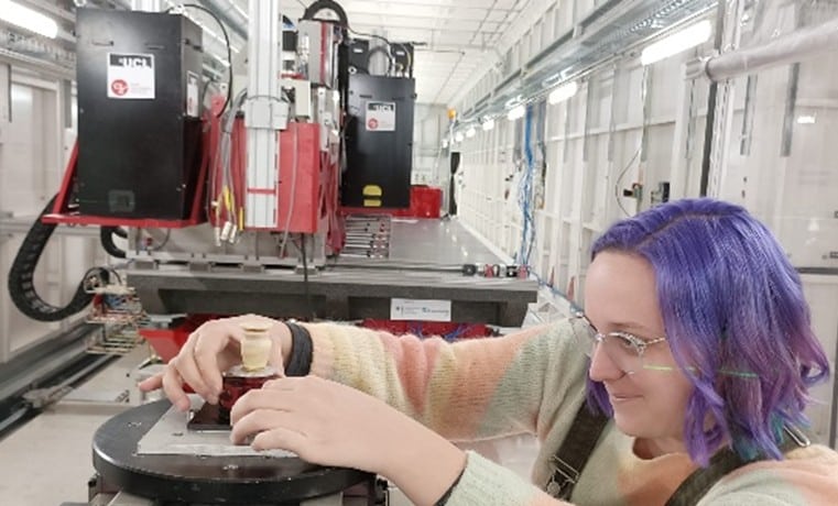
Synchrotron nanotomography: a unique technique only available at synchrotron radiation facilities
Atomic Force Microscopy (AFM)
Scanning Electron Microscopy (SEM)
Synchrotron microtomography (microCT)
Small-Angle X-ray Sacttering (SAXS)
A multidisciplinary team with deep expertise in analytical techniques used by scientists.
Cutting edge analytical approaches to support your product and process development, quality control, or marketing efforts.
Unique knowledge and expertise in analytical tecniques and 2D, 3D, and 4D imaging.