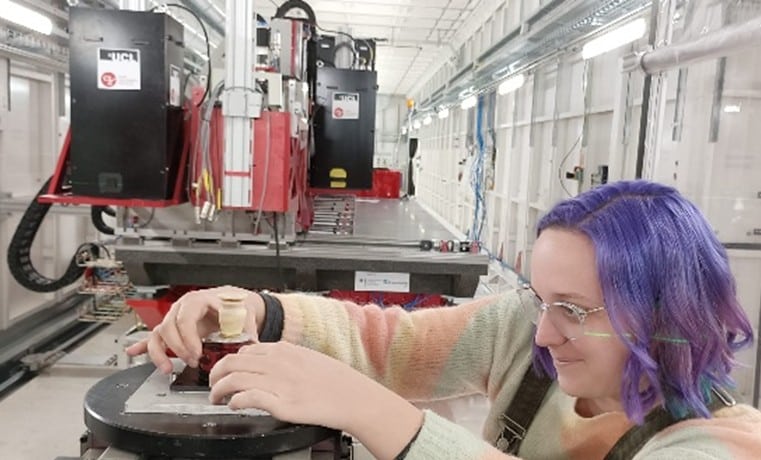
Tracking the evolution of materials microstructure : in situ mechanical testing with Novi CT Rig
Why perform microCT during tensile/compression tests? The use of…
Home » Technical means » X-ray tomography » In situ microtomography
In situ microtomography combines non-destructive 3D imaging (micro-CT) with the controlled application of stimuli (mechanical, thermal, chemical, electrochemical, moisture-related, etc.), allowing real-time observation of the internal evolution of a material or component. This transforms standard 3D imaging into a time-resolved 3D sequence under controlled conditions—hence the term “4D” or “in situ.”
The intensity and coherence of synchrotron radiation significantly enhance in situ tomography capabilities, enabling the tracking of material changes or deformations with unmatched speed and precision.

Ideal for fast phenomena, real-time monitoring, low contrasts, and high resolution.
Why perform microCT during tensile/compression tests? The use of…
Which phenomena are decisive in oral drug release from…
Why perform microCT during tensile/compression tests? The use of…
Which phenomena are decisive in oral drug release from…
3D analysis of porosity
3D analysis of fibres
3D chacracterisation of cracks
Advanced non-destructive imaging
Real-time and in situ tomography
Geometry comparison with CAD
Synchrotron analyses
Analysis of metals and alloys
Characterisation of composites and polymers
R&D support and partnership
A multidisciplinary team with deep expertise in analytical techniques used by scientists.
Cutting edge analytical approaches to support your product and process development, quality control, or marketing efforts.
Unique knowledge and expertise in analytical tecniques and 2D, 3D, and 4D imaging.