3D localisation and visualisation of cracks
Microtomography enables the non-destructive detection and 3D visualization of…
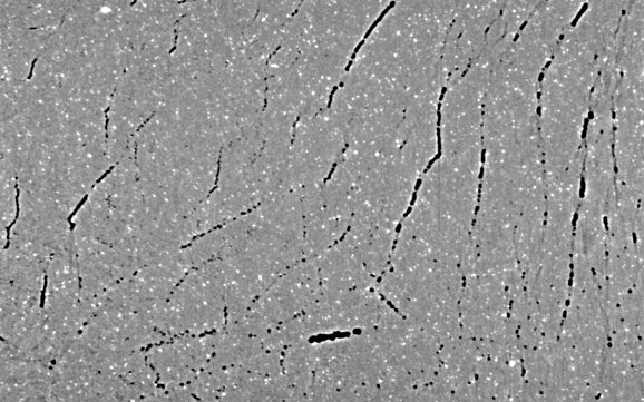

Microtomography enables the non-destructive detection and 3D visualization of…
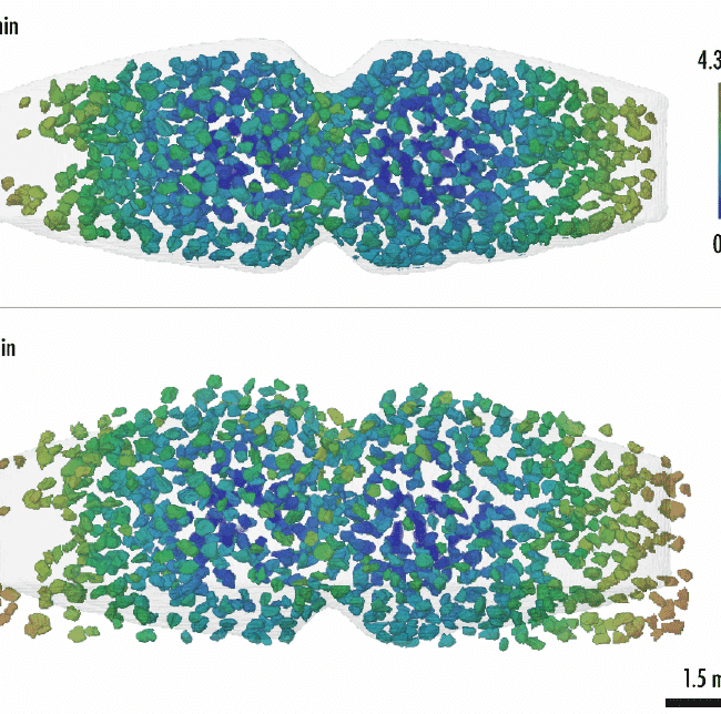
A recent study conducted by Xploraytion, Novitom, Merck and…

Why perform microCT during tensile/compression tests? The use of…
Microtomography enables the non-destructive detection and 3D visualization of…
A recent study conducted by Xploraytion, Novitom, Merck and…