Quantifying dermal collagen organisation with AI-assisted AFM
A new approach for assessing skin ageing and treatment…


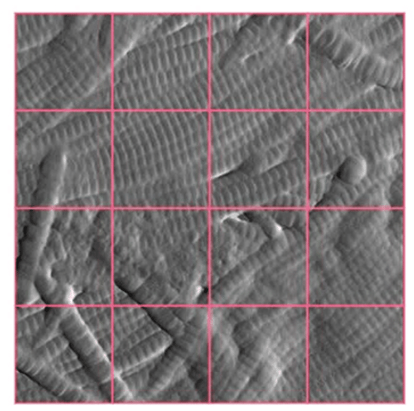
A new approach for assessing skin ageing and treatment…
X-ray tomography is now a reference method for non-destructive…

Why perform microCT during tensile/compression tests? The use of…
A new approach for assessing skin ageing and treatment…
X-ray tomography is now a reference method for non-destructive…