Agrifood

volume tomographique révélant d'intérieur d'une montre

Why choose X-ray tomography or microCT?

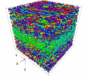
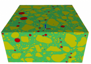
X-ray tomography is now a reference method for non-destructive three-dimensional imaging. Its principle is based on capturing a large number of X-ray images from different angles, which are then combined digitally to reconstruct the internal structure of an object. Tomography, which has become essential in various fields such as material

More »
visualisation 3D de l'orientation des fibres dans un polymère composite par tomographie RX

3D characterisation of fibres orientation in a composite

The images from X-ray microtomography (μCT) allow for advanced analyses at the fiber level: composites, fabrics, plants, paper. We have developed algorithms dedicated to high-resolution image analyses in order to calculate the orientation fields of fibers in samples or complete parts. The material is probed ‘as-is’, without the need to

More »
Volume 3D colorisé illustrant la segmentation des phases et grains dans du ciment.

Why choose microCT to study particle size?

X-ray microtomography (micro-CT) images enable advanced particle size analysis, allowing the characterisation of the size, shape, and spatial distribution of particles, pores, or inclusions that make up one or more phases of a material. The material is probed “as is” , i.e. without the need for conditioning. The three-dimensional nature

More »
extrait de webinaire décrivant l'insertion de défauts virtuels via le logiciel de simulation CT Novi-Sim

Optimizing X-ray CT with virtual tomography

On September 13, 2023, Rigaku hosted an episode of its Ask the Expert webinar series, featuring Dr. Awen Autret, R&D Engineer at Novitom. The session, titled “Virtual Tomography – Optimizing Data Acquisition Parameters Without a CT Scanner”, explored how advanced simulation can accelerate and enhance X-ray CT imaging workflows. Dr.

More »