Revealing the hidden structure of tablets with synchrotron X-rays
A recent study by Xploraytion, Novitom, Merck, and partners demonstrates how synchrotron X-ray microtomography (SR-µCT) is transforming the analysis of…
Accueil » Articles et actualités » Inclusions au sein de superalliages métalliques
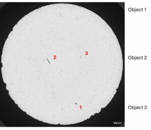
Afin d’étudier la rupture de pièces critiques comme peuvent l’être les aubes de turbine, la morphologie des inclusions de différents types au sein d’échantillons de superalliages métalliques à base nickel a été caractérisée : localisation, dimensions, formes, orientations. Les atouts de la microtomographie à rayons X synchrotron ont permis d’atteindre un contraste suffisant pour dissocier ces différentes inclusions, mais également des microporosités, d’être assez résolu (~1 µm) pour extraire des données quantitatives par analyse des images 3D.
What are the assets of synchrotron microtomography SR-μCT here?
++ Non-invasive, real-time analysis
SR-μCT allows direct, non-invasive monitoring of structural changes in pellets, in particular the formation of cracks in polymeric coatings, without complex sample preparation. This reduces the risk of creating artefacts and provides a more accurate analysis of the internal dynamics of solid pharmaceutical dosage forms.
++ Optimisation of formulations
By better understanding the underlying mechanisms of drug release, such as osmosis and mechanical stress, manufacturers can optimise the design of coated tablets, reducing the amount of trial and error usually required in the development of new formulations.
++ Improved modelling and prediction
The data obtained from SR-μCT is also used to develop models, making it easier to predict drug release behaviour as a function of the composition of coatings and the internal structure of tablets, lozenges or pills.
As an independent lab, Novitom enables companies to access synchrotron micro and nano-tomography :
A recent study by Xploraytion, Novitom, Merck, and partners demonstrates how synchrotron X-ray microtomography (SR-µCT) is transforming the analysis of…
Qui n’a jamais rêvé de voir à l’intérieur d’une montre sans avoir à le démonter pour observer ou contrôler la…
Les images issues de la microtomographie aux rayons X (µCT) permettent de réaliser des analyses avancées au niveau des fibres:…
A recent study by Xploraytion, Novitom, Merck, and partners…
Les images issues de la microtomographie aux rayons X…