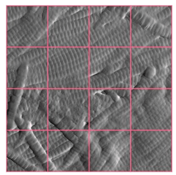
Anti-aging skincare seen through collagen fibres organisation
Novitom has developed an original test based on the…
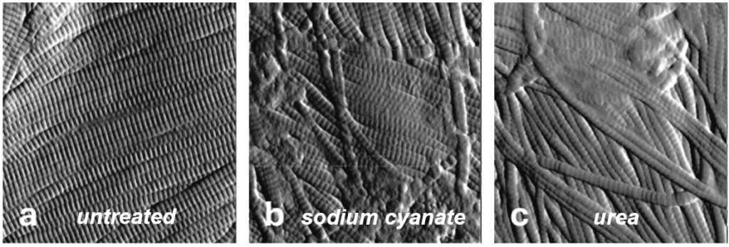

Novitom has developed an original test based on the…

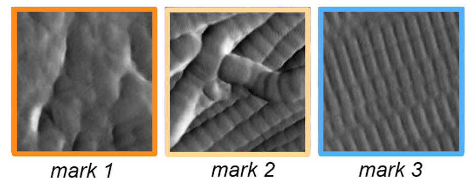
X-ray diffraction reveals the structural organisation of the intercorneocyte…