3D microscopy

View the microstructure in 3D without cutting

Novitom uses X-ray microtomography (micro-CT) to visualise and analyse materials and biological samples in three dimensions, without cutting or altering them.

This advanced imaging technique enables virtual histology, providing detailed information on microstructures. By combining high-resolution imaging and quantitative 3D analysis, we help researchers and engineers better understand the relationships between structure and function or mechanical and physical properties.

coupe tomographique d'une graine

Our expertise in 3D microscopy

  • Virtual histology
  • Visualisation and morphology of microporosities, lacunae, defects
  • Particle size analysis: ingredients, aggregates, inclusions, particles
  • Segmentation and quantification of different phases, fibres, ingredients, components, 
  • Analysis of textures and changes according to temperature and humidity
  • Surface tortuosity measurements or profilometry on virtual sections
  • In situ 3D microscopy: monitoring of microstructure evolution under mechanical stress

3D microscopy: a non-destructive technique to support your developments

  • Transformation of the microstructure of food products: extrusion, mixing, compression, humidification, pressure, cooking, deep-freezing, freeze-drying
  • Integrity and defects in packaging: delamination, microcracks, tears, sealing
  • Study of cellular structures: polymer foams, food foams
    3D and 4D visualisation of technical fabric fibres in a finished product

The advantages of NOVITOM

Reliable and rapid analyses

Analyses carried out on state-of-the-art equipment by a team of experts.

Tailor-made approach

An approach tailored to your constraints, with a technical contact person who supports you from start to finish.

Clear results

Actionable results thanks to detailed reports and 3D visuals that facilitate decision-making.

Further reading about 3D microscopy

Why choose microCT to study granulometry?

Why choose microCT to study granulometry?

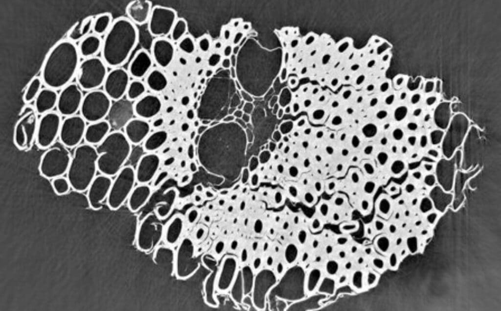
X-ray microtomography (micro-CT) images enable advanced particle size analysis,…

Revealing the hidden structure of tablets with synchrotron X-rays

Revealing the hidden structure of tablets with synchrotron X-rays

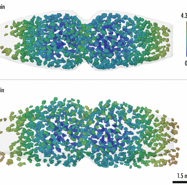
A recent study conducted by Xploraytion, Novitom, Merck, and…

Analyse et visualisation de volumes des glandes sébacées

Analyse et visualisation de volumes des glandes sébacées

La microtomographie à rayons-X, véritable microscopie 3D non destructive,…

Types d’échantillons

  • Matériaux biologiques d’origine végétale ou animale
  • Matériaux semi-solide ou visqueux
  • Matériaux fibreux
  • Matériaux solides : métaux, composites, matériaux compactés ou multicouches