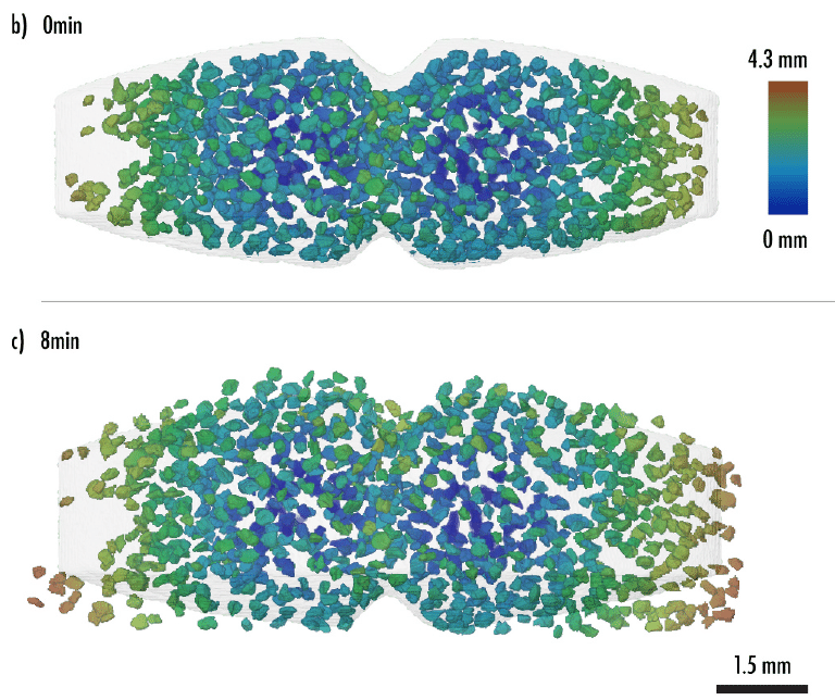
Revealing the hidden structure of tablets with synchrotron X-rays

A recent study conducted by Xploraytion, Novitom, Merck, and their partners demonstrates how synchrotron X-ray microtomography (SR-µCT) is transforming the analysis of pharmaceutical tablets. Published in the International Journal of Pharmaceutics, this study shows how the internal structures of tablets can be visualized in 3D and in real time.

The resolution, speed, and sensitivity of synchrotron X-rays make it possible to capture coating uniformity, porosity, and disintegration dynamics in just a few seconds.

A dissolution cell (developed by Xploraytion) enables realistic tablet disintegration studies under continuous or static flow conditions, paving the way for finer, non-destructive quality control and a better understanding of drug release mechanisms.

Original publication: Static and in-situ synchrotron X-ray microtomography analysis of pharmaceutical tablets – ScienceDirect

Context of the Study

This study demonstrates how synchrotron radiation microtomography (SR-µCT) overcomes these limitations by exploiting the exceptional properties of synchrotron X-rays—brightness, high coherence, and tunable energy—to achieve remarkable spatial and temporal resolution. This work presents both static and dynamic (or in situ) imaging of pharmaceutical tablets, offering new insights into coating integrity, internal porosity, and disintegration dynamics under realistic conditions.

This study demonstrates how synchrotron radiation-based microtomography (SR-µCT) overcomes these limitations by exploiting the exceptional properties of synchrotron X-rays – extreme brightness, high coherence, and tunable energy – to achieve outstanding spatial and temporal resolution. The work showcases both static and in-situ imaging of pharmaceutical tablets, offering new insights into coating integrity, internal porosity and disintegration dynamics under realistic conditions.

Static Analysis : investigation of tablet’s coating

A commercial pantoprazole tablet served as a model system for evaluating the trade-off between field of view (FOV) and resolution. Using voxel sizes ranging from 6.5 µm to 0.65 µm, SR-µCT visualised variations in coating thickness, pore networks, and sub-micron cracks that remain invisible in conventional µCT. The study showed that while low-resolution scans suffice for assessing global uniformity, high-resolution imaging is essential for detecting micro-defects and quantifying coating thickness.

The combination of phase contrast and high sensitivity enabled reliable identification of coating defects and porosity distributions, demonstrating that non-destructive 3D coating inspection of commercial tablets is now achievable with high accuracy.

Dynamic (In-situ) Analysis : investigation of API and cracks distribution during dissolution

A major technological advance presented in this work is a dissolution chamber. This custom-designed, fluid-circulating chamber enables in situ micro-computed tomography (MCCT) imaging of tablets during dissolution under real-world conditions. The chamber continuously circulates liquid from a 100 ml reservoir, ensuring homogeneous conditions. Its spring-loaded pressure system minimizes motion artifacts during rotation, a significant improvement over conventional static or syringe-based systems.

Using this device, the researchers performed time-resolved SR-µCT on placebo and commercial tablets (e.g., Beloc Zok) with acquisition times of 5 seconds per scan, capturing water penetration, swelling, and crack formation in real time. At a higher resolution (voxel size of 3 µm), this approach revealed a progressive fracture of the excipient phases and distinct double-layer coatings on the active ingredient granules, structural features impossible to resolve with laboratory instruments.

Thanks to the high photon flux of the synchrotron beam, the team could continuously track dissolution behaviour over time, eliminating the need to sacrifice multiple samples at different time points. This represents a step change towards true four-dimensional imaging of drug release mechanisms.

Unique Capabilities of Synchrotron X-rays for the study of pharmaceutical tablets

Compared to laboratory X-ray sources, synchrotron radiation provides X-ray beams that offer the following characteristics for studying solid pharmaceutical dosage forms:

  • Submicron spatial resolution, down to a few hundred nanometers;
  • Phase-contrast imaging, which reveals materials with small density differences, such as polymer coatings or excipients;
  • Ultrafast acquisition, enabling the capture of complete 3D tomograms in seconds rather than minutes or hours;
  • Time-resolved (4D) imaging, recording structural changes during swelling or dissolution;
  • Large fields of view thanks to specific acquisition geometries, allowing for highly detailed images of entire tablets.

As an independent lab, Novitom enables organisations to access synchrotron micro and nano-tomography :

  • Regular access to instruments at synchrotron facilities
  • Know-how in experimental design for real-time microtomography
  • Expertise in extracting information and physical parameters from raw data and in combination with ancillary techniques