Tracking the evolution of materials microstructure : in situ mechanical testing with Novi CT Rig
Why perform microCT during tensile/compression tests? The use of…

Why perform microCT during tensile/compression tests? The use of…
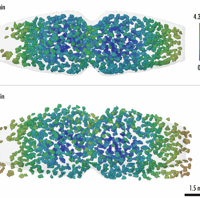
A recent study conducted by Xploraytion, Novitom, Merck and…