In situ microtomography

In situ microtomography combines non-destructive 3D imaging (micro-CT) with the controlled application of stimuli (mechanical, thermal, chemical, electrochemical, moisture-related, etc.), allowing real-time observation of the internal evolution of a material or component. This transforms standard 3D imaging into a time-resolved 3D sequence under controlled conditions—hence the term “4D” or “in situ.”

The intensity and coherence of synchrotron radiation significantly enhance in situ tomography capabilities, enabling the tracking of material changes or deformations with unmatched speed and precision.
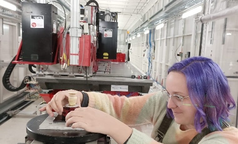
Advantages of In Situ Tomography

  • Time-resolved monitoring and non-destructive 3D imaging in a controlled environment
  • Wide range of compatible equipment: tensile/compression machines, furnaces, climate chambers, cooling systems
  • Fast acquisition mode for studying rapid events in real time: cracking, dendrite formation, wet granulation, delamination
  • Adaptable image resolution: pixel size from approximately 0.5 µm to 50 µm
  • Unique technology for digitizing processes
  • Multi-field experiments possible, combining heating, mechanical loading, controlled atmosphere, electrical/electrochemical input (in operando), to closely replicate real-world conditions
  • Suitable for all types of materials

Measurements and Analyses using real-time microtomography

  • Mechanical testing: tensile/compression tests on specimens
  • Monitoring of sintering and densification processes
  • In situ thermal testing: temperature cycles, thermal shocks
  • In operando electrochemical experiments on batteries and fuel cells
  • In situ corrosion / chemical reactions: deposit growth, chemical degradation
  • Monitoring of tablet dissolution 
  • Digitization of food processes: cooking, freeze-drying, hydration, emulsions
  • Custom design of experiments or stimulation equipment

Novitom's Expertise in real-time tomography

  • Custom design of stress equipment
  • Tailored preparation of tests and samples for efficient use of X-ray beam time
  • Optimized imaging protocols, selected based on your specific needs: quasi-static or dynamic monitoring, high resolution, strong contrast
  • Advanced 3D data processing: segmentation, pre-trained AI networks, DVC (Digital Volume Correlation), quantitative measurements, interpretation
  • Directly usable results: provided as reports or digital data compatible with your software
  • Comprehensive support: from defining your needs to final delivery, backed by over 20 years of experience

Synchrotron tomography vs. laboratory µCT for in situ studies
When to choose which?

In situ tomography (laboratory-based source)

  • Ease of access for slow dynamics (over several days or weeks)
  • Scan rate limited by the power of the source
  • Pixel size approximately ≥ 5 µm to limit scan times
  • Absorption contrast
  • Space constraints due to the cabin dimensions
 
 Ideal for slow phenomena, quasi-static monitoring, intermediate resolutions, and for preparing synchrotron measurements.

In situ synchrotron tomography

  • Very high scan rates (< 1 second per 3D scan)
  • High resolution available (< 1 µm)
  • Phase contrast, low noise
  • Few space constraints for installing in situ devices
  • Limited suitability for slow dynamics
  •  

Ideal for fast phenomena, real-time monitoring, low contrasts, and high resolution.

Types of sample

  • Metals and Alloys: Specimens, coupons, lattice structures, powders, coatings
  • Polymers and Composites: Carbon/carbon, SMC (Sheet Molding Compound), 3D woven, CMC (Ceramic Matrix Composites), polymer-metal, foams
  • Ceramics and Glasses: Dense or porous samples, powders
  • Pharmaceutical and Cosmetic Products: Tablets, gels, emulsions
  • Batteries, Electrodes, Membranes: In operando monitoring
  • Biological Tissues: Bones, teeth, soft tissues, plant and food samples

Examples of analyses and tests using in situ tomography

To stress materials and products while imaging them via X-ray or synchrotron microtomography, we design miniaturised devices that meet the requirements of both testing and 3D imaging.

  • Tensile/compression machines: from 500 N to 20 kN
  • Heating stages / laser heating: from room temperature to >1000–2000 °C for high-temperature testing
  • Climatic chambers: with controlled humidity (10% RH – 90% RH); temperature range from -40 °C to +60 °C
  • Cryogenics
  • Sample changers: for in operando electrochemical cell testing
  • Fluid injection chambers: for liquids and corrosive atmospheres

Complementarity with other techniques

Microtomographie rayons-X in situ 

Microtomographie rayons-X (µCT)

Microscopie électronique à balayage basse tension (SEM-FEG)

Microscopie à force atomique (AFM)

Nanotomographie synchrotron

Fluorescence rayons-X en microfaisceaux (µXRF)

Micro-diffraction à rayons X

NOVITOM: Your Partner in R&D, Imaging, and Advanced Analysis

Expertise

A multidisciplinary team with deep expertise in analytical techniques used by scientists.

Innovation

Cutting edge analytical approaches to support your product and process development, quality control, or marketing efforts.

Support

Unique knowledge and expertise in analytical tecniques and 2D, 3D, and 4D imaging.